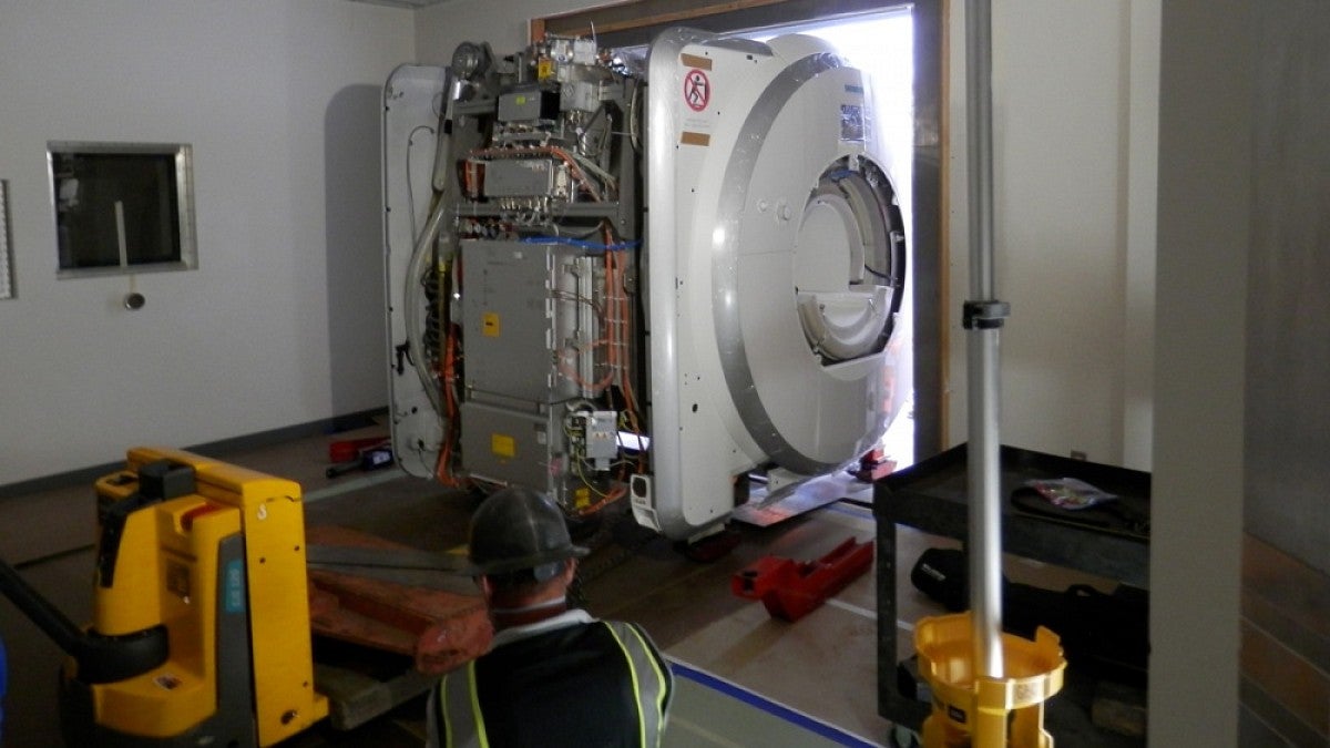
Workers hoisted a new 13-ton magnetic resonance imaging scanner into the Lewis Integrative Science Building last week, doubling the capacity of the UO’s advanced imaging lab.
The new Siemens Prisma scanner is part of the Robert and Beverly Lewis Center for Neuroimaging.
“Due to the tremendous support from the Lewis family, our researchers will have unparalleled access to imaging instruments, and the new Prisma scanner will produce better scans, improving the impact of the science our faculty members are conducting,” said Fred Sabb, director of the center.
Magnetic resonance imaging, a noninvasive imaging technology that produces three-dimensional, detailed anatomical images, can be used to tell the difference between various types of tissues. It is often used in medical settings for disease detection, diagnosis and treatment monitoring.
Research-dedicated instruments such as those at the Lewis Center for Neuroimaging have specialized hardware and software to conduct functional magnetic resonance imaging that can be used to observe brain function in near real time by observing which areas of the brain consume more oxygen during various cognitive tasks, suggesting they are working harder.
Researchers in the Lewis Center for Neuroimaging use MRIs in studies examining everything from energy production in muscles to brain activity in teenagers. UO researchers with expertise in psychology, neuroscience, human physiology and an array of other disciplines conduct research at the center.
The new MRI is being installed in a former conference room converted into an MRI bay. Widely considered the best research magnet currently available, the Prisma is used by the majority of top-tier research institutions engaged in brain imaging, Sabb said.
It will allow the UO to handle a growing number of researchers conducting studies involving MRI technology. The high demand is partly due to the recent hiring of new faculty members in the Department of Psychology, Department of Human Physiology and College of Education. The center will continue to operate its existing MRI, a Siemens Skyra acquired in 2012.
The new scanner will also allow greater collaboration with researchers at Oregon Health & Science University, which has an identical instrument. It will enable investigators to conduct research in tandem at both institutions and could lead to the UO’s involvement in more ambitious research projects.
“We are talking to OHSU and other institutions about partnering on larger grant applications, which are the types of studies that federal funding agencies have been prioritizing in our field,” Sabb said. “This has already opened up significant doors for us in that area, and it’s a tremendous asset for our faculty and students that represents a real competitive advantage for us.”
The acquisition of the new MRI was funded by the Robert and Beverly Lewis Center for Neuroimaging Endowment Fund, established in 2001, with additional support provided by the Office of the Vice President for Research and Innovation. It is expected to be fully operational in mid-August.


