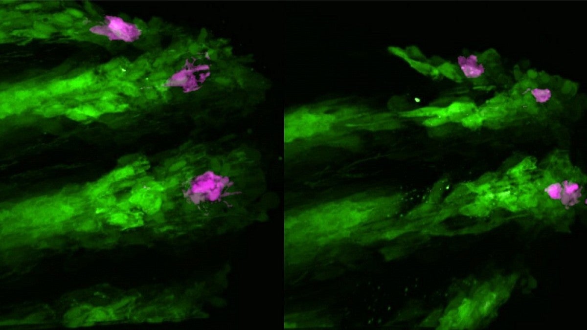
A new high-tech microscope is the latest in a long line of advanced instruments headed to UO labs thanks to the generous support of the M.J. Murdock Charitable Trust.
The trust, based in Vancouver, Washington, has provided almost $10 million since its inception in 1975 to support research and discovery at the university. Its latest contribution to UO science, which is due to arrive this summer, is a spinning disc confocal microscope.
“I think it’s going to be really useful to a lot of people,” said molecular biologist Brad Nolen. “This is going to have a significant impact on the science we can do here.”
The microscope will be the go-to instrument for researchers like Nolen, who will use it to perform live cell imaging in multiple dimensions. It should arrive on campus early this summer, thanks to a new instrumentation grant from the trust.
Nolen, an associate professor in the Department of Chemistry and Biochemistry and the Institute of Molecular Biology, played a key role in securing the grant.
“In labs, research centers and core facilities across campus, you can see the enormous impact of the M.J. Murdock Charitable Trust’s many contributions,” said David Conover, vice president for research and innovation. “They have boosted our research capacity in incalculable ways, and we are forever grateful to them for their continued support of the UO’s research mission.”
Founded in 1975 at the bequest of Melvin J. Murdock, the trust set out to enrich the quality of life in the Pacific Northwest. For the region’s universities, this translated to investments in research infrastructure, education and cultural programs.
At the UO, the support has enabled such diverse research as functional MRI studies on the link between brain activity and behavior, genomic analysis to identify molecular signatures of cells, and fabrication of thin-film devices with applications in energy, communications and health.
The new microscope will be housed in the UO’s Genomics and Cell Characterization Core Facility, a core research facility that was recently renovated thanks to a $10 million gift from Mary and Tim Boyle.
The powerful imaging tool will fill a gap in the facility’s capabilities and will help open new doors of discovery for researchers. In addition to providing the highest-resolution imaging currently available, it will allow researchers to probe and measure biomolecules and perform other operations that can help explain how molecules are functioning within a cell, Nolen said.
One of the ways Nolen will use the instrument will be to tag proteins within cells so he can control and observe their interactions, which could help further his understanding of how proteins are involved in particular biological functions.
Researchers in his lab study actin, a protein that gives cells structure and the ability to move and interact with other cells. Because actin is involved in many processes that occur in healthy cells, understanding its function can shed light on bacterial infections, viral infections and other disease states where cells are not healthy, Nolen said.
“The microscope will enable students and faculty to develop innovative methodologies in their pursuit of such fundamental questions as how proteins and other biomolecules drive biological function in healthy and diseased cells,” said Moses Lee, senior director for scientific research and enrichment programs of the M.J. Murdock Charitable Trust.


