
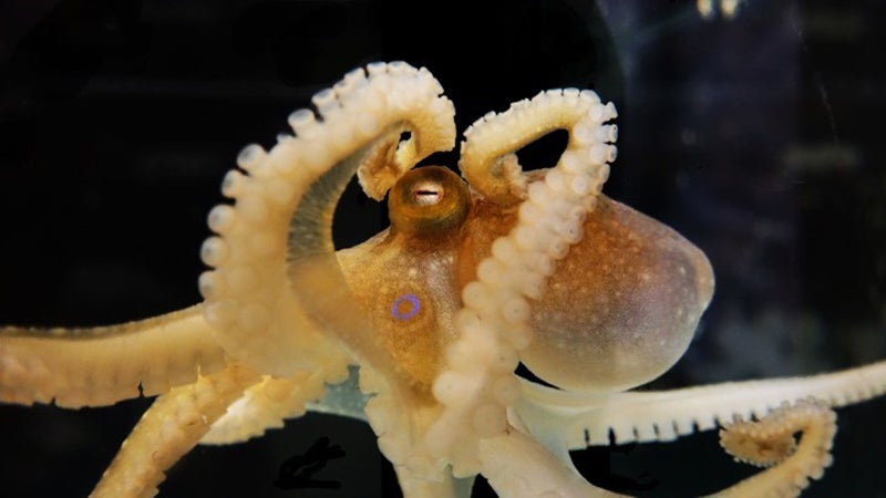
An octopus devotes about 70 percent of its brain to vision. But until recently, scientists have only had a murky understanding of how these marine animals see their underwater world. A new University of Oregon study brings the octopus’ view into focus.
For the first time, neuroscientists have recorded neural activity from the visual system of an octopus. They’ve created a map of the octopus’ visual field by directly observing neural activity in the animal’s brain in response to light and dark spots in different locations.
This map of the neural activity in the octopus visual system looks a lot like what’s seen in the human brain, even though octopuses and humans last shared a common ancestor some 500 million years ago, and octopuses have evolved their complex nervous systems independently.
UO neuroscientist Cristopher Niell and his team in the UO’s College of Arts and Sciences report their findings in a paper recently published in Current Biology.
“Nobody has actually recorded from the central visual system of a cephalopod before,” Niell said.
Octopuses and other cephalopods aren’t typically used as a model for understanding vision, but Niell’s team is fascinated by their unusual brains. In a related paper published last year in Current Biology, the lab identified different categories of neurons in the octopus optic lobe, the part of the brain dedicated to vision.
Together, “these papers provide a nice foundation by elucidating the different types of neurons and what they respond to — two essential aspects we’d want to know to start understanding a novel visual system,” Niell said.
In the new study, researchers measured how neurons in the octopus visual system responded to dark and light spots moving across a screen. Using fluorescent microscopy, the researchers could watch the activity of neurons as they responded, to see how neurons reacted differently depending on where the spots appeared.
“We were able to see that each location in the optic lobe responded to one location on the screen in front of the animal,” Niell said. “If we moved a spot over, the response moved over in the brain.”
This kind of one-to-one map is found in the human brain for multiple senses, like vision and touch. Neuroscientists have connected the location of particular sensations to specific spots in the brain.
A well-known representation for touch is the homunculus, a cartoon human figure in which body parts are drawn in proportion with how much brain space is devoted to processing sensory input there. Highly sensitive spots like fingers and toes appear massive because there are lots of brain inputs from these body parts, while less sensitive areas are much smaller.
But finding an orderly linkage between the visual scene and the octopus brain was far from a given. It’s a fairly complex evolutionary innovation, and some animals, such as reptiles, don’t have that kind of map. Also, previous studies had suggested the that octopuses do not have a homunculus-like map for different parts of their body.
“We hoped that the visual map might be there, but nobody had directly observed it before,” Niell said.
The octopus neurons also responded particularly strongly to small light spots and big dark spots, the researchers noted, a distinct difference from the human visual system. Niell’s team hypothesizes that this might be due to the specific characteristics of the underwater environment that octopuses must navigate. Looming predators might appear as large dark shadows, while close-up objects like food would appear as small bright spots.
Next, the researchers hope to understand how the octopus brain responds to more complex images, such as those actually encountered in their natural environment. Their eventual goal is to trace the path of these visual inputs deeper into the octopus brain, to understand how the octopus sees and interacts with its world.
—By Laurel Hamers, University Communications