
Studying sex from just the male perspective misses half the experience. But much of what we know about the origins of reproductive cells comes from looking at sperm and egg formation separately — or only focusing on sperm.
Now, new University of Oregon research in tiny worms is directly comparing the two processes, unpacking some of the ways that the same building blocks get used differently to make sperm and eggs. A team from the lab of UO biologist Diana Libuda in the College of Arts and Sciences reports their findings in a paper published Oct. 5 in the journal eLife.
“This study starts to open the door to understanding these sex-specific differences,” said Cori Cahoon, a postdoctoral fellow in Libuda’s lab who led the work.
Understanding how these processes work normally can help researchers figure out what’s gone wrong in cases of infertility.
In mammals, it’s hard to study egg and sperm development side by side. Sperm are made continuously throughout an animal’s lifespan, but the process of egg formation starts when the developing animal is still in utero.
“There's a very narrow window in human development when egg formation happens,” Libuda said. “It’s a very hard window to catch.”
Not so in C. elegans, a species of small worm that’s commonly used for genetics research. Most of the proteins involved in worm fertility are also found in humans. But C. elegans make egg and sperm continuously throughout their lifespan, making it easier to directly compare the two processes.
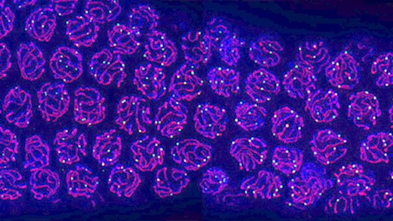
Plus, worms devote about three-quarters of their body to making sperm and eggs. And the tiny critters are transparent, so the process is clearly visible under the microscope.
To watch sperm and eggs develop, Cahoon developed an imaging technique to immobilize worms under the microscope. She used a gene that controls the worms’ movement and bred the worms so the gene stops working in the presence of a plant hormone.
When researchers put the worms under the microscope on petri dishes coated in this hormone, the worms stood still for their portrait. But they also recovered movement quickly as they were removed from the hormone-coated plates. That simple technique allows researchers to visualize live egg and sperm development throughout the animal’s lifespan.
Both eggs and sperm are made via meiosis, a multistep process that evenly divides a cell’s genetic material to create reproductive cells with the correct number of chromosomes. While the big picture steps are the same regardless of sex, small differences add up to yield two very different products.
For example, during meiosis chromosomes link together and swap pieces, mixing up genes.
This step, called recombination, is important for generating genetic diversity and ensuring the correct number of chromosomes are inherited.
Recombination happens at a different rate in eggs compared to sperm, past research has shown. And in sperm, the process is highly sensitive to temperature; in eggs, it’s less so. But it wasn’t clear what was causing those differences.
In the study, the researchers specifically looked at a structure that forms during recombination and acts like a brace, holding the chromosomes in place as they swap pieces.
The same proteins are involved in making that supporting structure in eggs and sperm. But they’re used in different quantities, the team found, like using the same Lego kit to build two different airplanes.
That affects the way the structure holds the chromosomes together, helping to explain the different recombination rates. It might also help explain why the brace-like structure falls apart under high temperatures in developing sperm, but not in eggs.
Understanding more about how temperature affects reproductive cell formation is a major next step for the researchers.
“This research brings to light the importance of doing sex-comparative studies of sperm and egg development,” Libuda said. “You can start to illuminate special features that were previously overlooked, and really understand what the key players and differences are for sperm and egg development.”
The research was funded in part by the National Institutes of Health.
—By Laurel Hamers, University Communications
—Top photo: A magnified view of eggs developing in a model organism (Photo by Cori Cahoon)