
It’s now easier than ever to visualize the tiny building blocks of our world, thanks to the efforts of a University of Oregon physicist.
Kayla Nguyen has co-led the development of a new approach that allows scientists to see individual atoms and the way they fit together under an electron microscope, without the multimillion-dollar price tag that such ability typically commands.
The latest step in her ongoing efforts to make high-resolution electron microscopes more affordable and accessible is described in the Feb. 22 issue of the journal Science. Nguyen co-led the recent work as a postdoctoral researcher in the lab of Pinshane Huang at the University of Illinois. And the advance relies on a tool that Nguyen co-invented as a doctoral student at Cornell University, for which she won the Lemelson-MIT Prize for student inventors.
“People in the last hundred years have been building better and better lenses to look at things that are increasingly small, but there’s a huge barrier to affording the infrastructure that allows you to work at this scale,” Nguyen said. “My work shows that you can image very small atoms without having to spend the upfront cost for an expensive microscope.”
Scientists in research labs around the world depend on electron microscopes for high-resolution imaging. The best of those microscopes can reveal details that once seemed impossibly small; not just individual atoms, but even the spaces between them.
Even tiny changes to the structure of a material can have a big impact on its properties. So electron microscopes are valuable tools for designing new kinds of materials for clean energy, drug delivery, computing technology and many other applications.
But currently, this subatomic resolution is only possible with a top-tier microscope with an expensive lens. The package can run more than $3 million, putting it out of reach for many universities. In their recent Science paper, Nguyen and her colleagues describe a different way to get similar resolution at a much lower cost, using the power of physics.
Electron microscopes use a beam of electrons to probe the surface of a material. As electrons hit the material, they bounce off and hit a detector. The way that the material deflects the electrons, and thus the pattern those electrons make on the detector, reflects the material’s structure.Using a powerful microscope with an expensive lens, you can use a very focused electron beam. That will create a higher-resolution image, like drawing with a fine-line pen compared to a chunky marker.
But there’s another way to improve resolution, too — via the detector itself. The information collected by the detector is the same whether you’re using the fine line pen beam or the thick marker beam of a less fancy scope, Nguyen said. So if you can find a way to get more information out of the detector, you can improve the image quality even if you’re using a lower-resolution microscope.
She compares a standard detector to a low-quality cell phone photo where the bright sun gets washed out, but the person in the foreground is shadowed; it gets saturated easily, and so can’t accurately capture all the information in the scene. The detector that Nguyen helped develop as a graduate student, called the EMPAD detector, can gather information from millions of electrons at once without getting saturated.
Paired with the improved detector, Nguyen and her colleagues used a technique called ptychography. That approach uses a series of mathematical equations to piece together a high-resolution image by scanning the same sample many different times from slightly different positions. Ptychography removes distortions in the image via calculations, instead of using a lens.
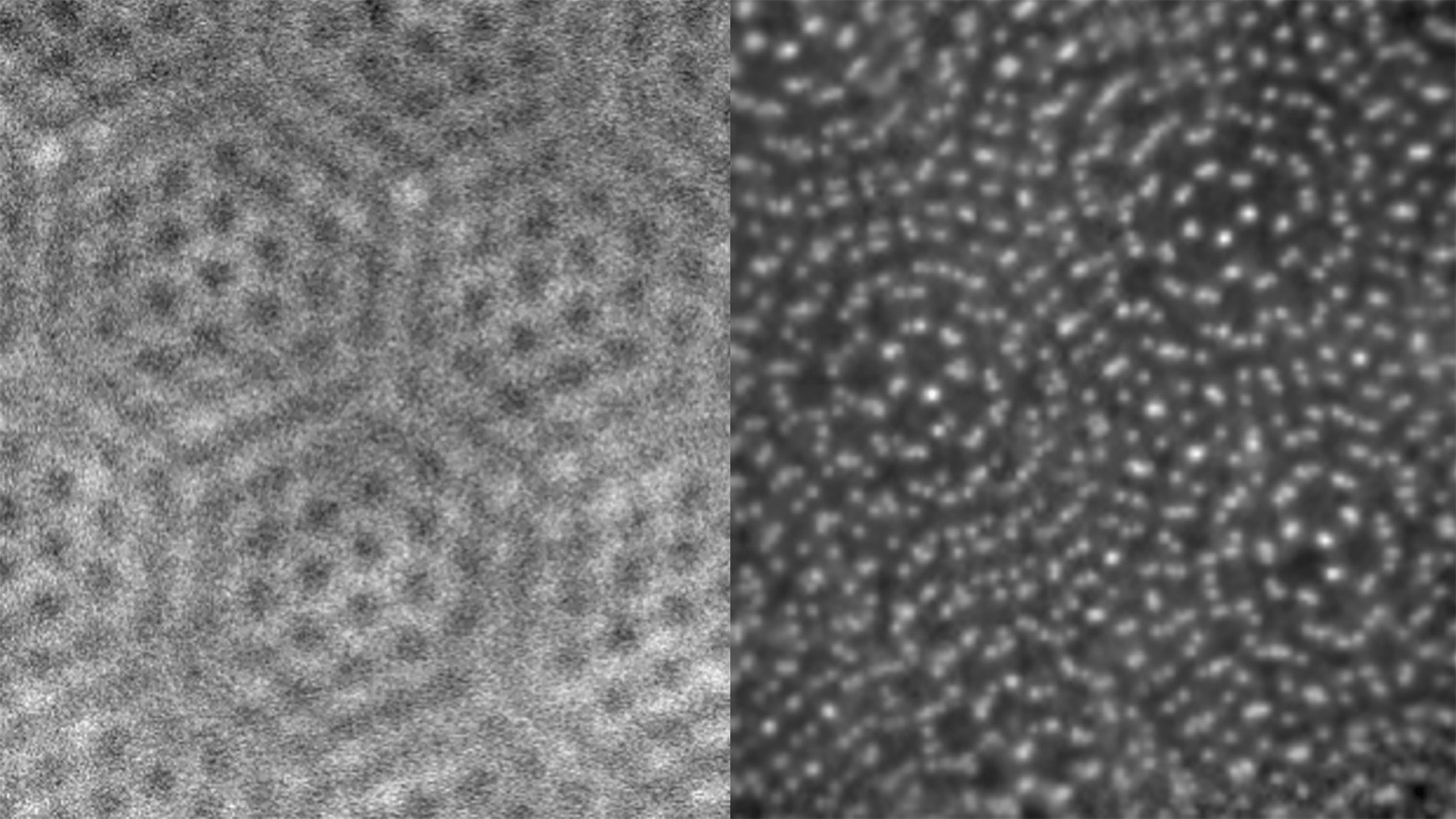
Combining the power of ptychography with the improved detector can turn an average-quality electron microscope into one that’s on par for resolution with the most state-of-the-art machines available, Nguyen and her colleagues found. In their new paper, they apply the technique to a twisted 2D material: two flat sheets with a small gap between them. Both the structure of the sheets and the gap between them was visible under the scope.
While others have used this improved more technique with an expensive microscope with good results, “we did it with a cheap microscope and a powerful detector,” Nguyen said.
The team tested the approach on a readily available commercial electron microscope, compared to a specialty scope that costs more than $3 million. The detector, which is licensed to Thermo Fisher Scientific, runs about $400,000. That puts it in reach of far more labs, expanding the potential for advanced imaging techniques.
As she sets up her physics lab in the UO's College of Arts and Sciences, Nguyen hopes to take the techniques even further and continue to refine the approach. She’s interested in diving into new kinds of advanced imaging and continuing to show that you don’t need the most expensive technology to get high-quality data and make meaningful contributions to science.
“Getting the highest-quality data doesn’t require the highest-resolution equipment, if you understand the physics to push it in different ways,” Nguyen said.
—By Laurel Hamers, University Communications
—Photo: The image on the left was made with a conventional electron microscope, while the one on the right shows the enhanced resolution from the new technique. (Image: Kayla Nguyen Lab)