
University of Oregon researchers are filling in gaps in our understanding of the formation and growth the colon, a crucial part of the digestive system, by being among the first to apply modern molecular biology tools to the study.
Understanding how a healthy colon forms could provide the key to treating common diseases, from inflammatory bowel disease to colon cancer. Yet some of the last significant research on colon development was conducted 30 years ago, before the current era of molecular biology.
A UO study, recently published in the American Journal of Physiology-Gastrointestinal and Liver Physiology, picks up where that research left off, applying the latest molecular and genetic tools to construct a step-by-step map of how colon tissue builds itself, using mice as a model organism.
The study is one of the first to define the structural and molecular characteristics of the developing mouse colon three weeks after birth.
Using fluorescent markers to track the activity of stem cells, the body’s building blocks, doctoral researcher Rachel Hopton investigated the formation of colon tissue in mice, which have long been used as a model for studying the gastrointestinal system in mammals. Because mice are born without fully developed colons, she was able to observe the process from birth to 21 days to provide new insight into the early stages of colonic development.
“A piece of the puzzle has been missing,” Hopton said. “We wanted to map out how the colon develops and over what time frame. No one has ever done that before.”
Since the 1990s, most gastrointestinal biologists have focused on the small intestine, which is more accessible and easier to study than the colon, which is sometimes called the large intestine. But although the two organs are molecularly similar in many ways, they have different structures and roles within the body, said biology professor Annie Zemper in the UO’s College of Arts and Sciences.
“In the world of biomedical research, it’s really rare to have something that hasn’t been looked at in such detail before, particularly in a common organ like the colon,” said Zemper, who directed the study along with co-author and lab technician Nicholas Jahahn. “By taking a whole-animal physiological approach, going step by step, we can build a model of how the colon constructs itself.”
Hopton also examined the role a protein known as Lrig1 plays in colon development. The protein, which is present in many human organs from the brain to the skin, acts as a tumor suppressor in adults by preventing the over proliferation of cells. By removing Lrig1 from some of the mice, she discovered that, unlike an adult colon, the developing colon was able to partially compensate for the loss.
“If you can understand some of the mechanisms of how the organ develops, you’ll understand what is happening during the process of disease and when that organ is breaking down,” she said. “You can’t figure out what’s going wrong until you know what has to be there in the first place. If we know how the colon develops, we can potentially use this information to make more informed decisions when treating gastrointestinal diseases.”
Because this was the first molecular study of its kind, Hopton had to devise many of her own research techniques. For example, she adapted a genetic cell-labeling process known as “Brainbow,” used by neuroscientists for distinguishing different neurons in the brain, to mark stem cell populations in the colon with multicolor fluorescent dyes to track their behavior over time.
“I had to pioneer everything I was doing from scratch, including how to handle this tissue at these earlier timepoints,” Hopton said. “The colon is so, so small compared to the small intestine, and it’s fragile. How do you handle it without compromising the sample? The whole first year was trial and error.”
Hopton’s study paves the way for further research, raising questions about how cells work together to form such a complex organ, how the microenvironment around the colon helps support its development, and what the long-term impacts of losing the Lrig1 protein are.
"We’re laying the groundwork for more mechanistic studies into this fascinating organ in the future,” said Hopton, who has since finished her doctorate and is now conducting gut-brain research for the U.S. Air Force to help improve human performance.
“I think it’s going to be interesting to see where the field goes after this,” she said. “Hopefully this will pique people’s interest to go back and do more developmentally focused colonic studies.”
—By Nicole Krueger, College of Arts and Sciences
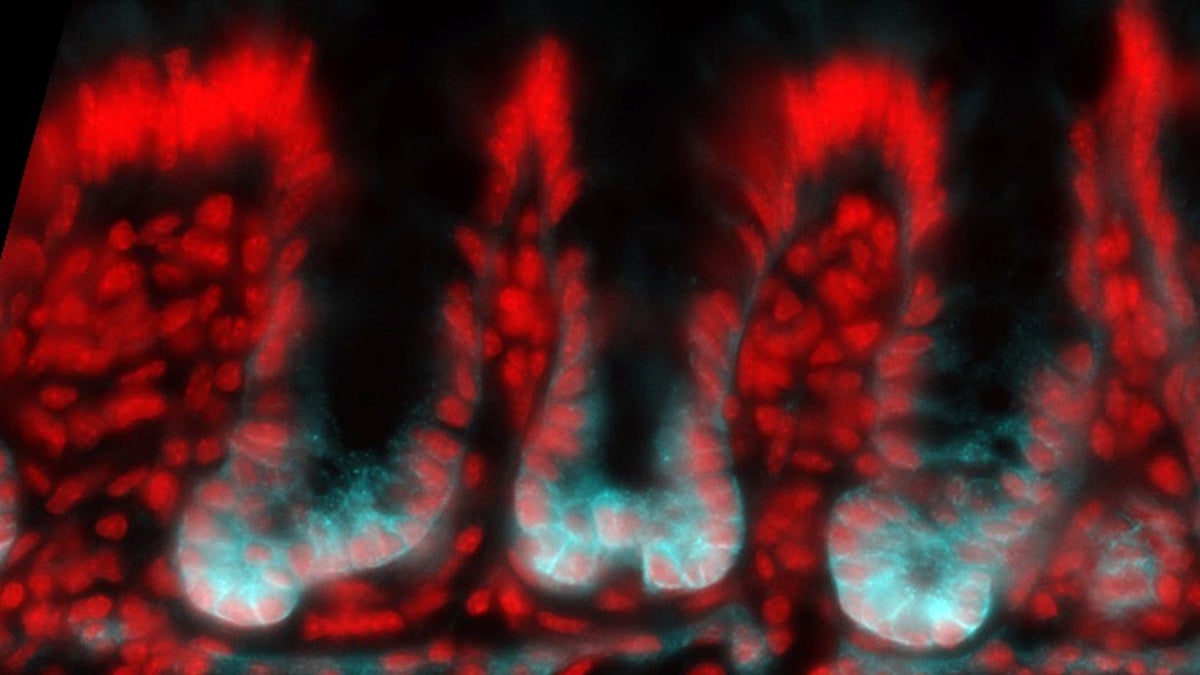
—Top photo: Epifluorescence image of the developing mouse colon from the study. All cells of the colon lining are bright red, while the specialized stem cells are also bright blue.